Immunofluorescence Cases
Georgia Dermatopathology Associates is pleased to offer a state-of-the-art immunodermatology service.
We offer both direct and indirect immunofluorescence (DIF, IIF) studies with multiple, simultaneous fluorochromes, DNA nuclear counterstains and other counterstains, and salt split skin analysis. We also apply additional antigen characterization via immunoblotting(IB) and ELISA techniques in appropriate cases. All case data is H&E correlated in one report, and signed out by a combined Dermatopathologist MD/Dermatoimmunologist MD, PhD team with over twenty combined years of independent signout experience.
Direct and Indirect Immunofluorescence
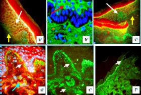
Figure legend: a and c. Direct immunofluorescence(DIF), demonstrating intercellular staining between epidermal keratinocytes using fluorescein isothiocyanate (FITC) conjugated anti-human IgG (green staining; white arrows); in addition, positive staining at the epidermal basement membrane zone (linear green staining; yellow arrows) is present, secondary to FITC conjugated Complement/C3 (linear yellow staining; yellow arrows); these images represent analysis of a biopsy from a patient affected by with pemphigus erythematosus (Senear-Usher syndrome). Note in c, the skin is folded near the bottom center, creating a visual artifact. The epidermal keratinocyte nuclei in a and c were counterstained with TO-PRO®-3/DNA(red staining). b, Indirect immunofluorescence (IIF) using FITC conjugated anti-human IgG, demonstrating positive staining to epidermal stratum corneum keratinocyte cytoplasms in a patient with active plaque psoriasis (green staining; red arrow). In addition, the keratinocyte nuclei were counterstained with 4',6-diamidino-2-phenylindole (Dapi; blue staining).d and e. DIF demonstrating positive staining at the epidermal basement membrane zone in a dermatitis herpetiformis(DH) patient biopsy, utilizing FITC conjugated anti-human IgA (yellow/green staining; upper white arrows). Note the classic, “snow on the mountains” pattern suprajacent to the dermal papillary tips. In addition, the light blue arrow in d and lower white arrow in e highlight circular, DH bodies in the superficial papillary dermis. In d, the red staining in the epidermis and dermis represent positive immunoreactivity with Texas red conjugated antibody to armadillo repeat gene protein deleted in velo-cardio-facial syndrome (ARCVF). ARVCF represents a catenin protein integral to cellular adherens junctions, and is also utilized in GDA research applications. In d, the epidermal keratinocyte nuclei were also counterstained Dapi(blue). f. IIF, created with normal human skin substrate and serum from an epidermolysis bullosa acquistia(EBA) patient, including salt-split treatment with 1 M NaCl. The IIF demonstrates deposits of FITC conjugated Complement/C3 at the epidermal basement membrane zone blister floor(linear green staining; white arrow).