Immunohistochemistry
Georgia Dermatopathology Associates offers an in-house, comprehensive selection of immunohistochemistry (IHC) antibody stains.
Providing:
- correlative excellence in H&E and DIF/IIF diagnoses
- improved diagnostic turnaround time
- investigative power in dermatology research applications
Below are examples of our work.
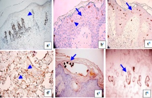
Figure legend: a. Myeloid/histoid antigen staining on neutrophils clustered within the dermal papillae, from a patient with dermatitis herpetiformis (brown staining; blue arrow). b, c, d Patient with early bullous spongiotic dermatitis. b. Specific, CD1a positive staining on epidermal Langerhans cells (blue arrows). c. Sensitive, S-100 staining on melanocytes, and on the same Langerhans cells as in b (blue arrow). d. Factor XIIIa positive, enlarged dendrocytes in the reticular dermis (blue arrows). e. Positive staining for herpes simplex virus 1(HSV-1) inside a patient’s facial epidermal blister (blue arrow). f. Complement 5b-9/MAC complex antibody, demonstrating delicate, focally positive dermal eccrine gland staining in a dermatomyositis patient (blue arrow)