Bullous Allergic Contact Dermatitis
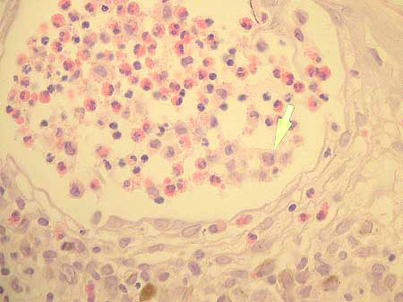
Microphotograph Series DSCN0161Intraepidermal vesicle featuring numerous lymphocytes, eosinophils and Langerhans histiocytic cells(Arrow), H&E 400X
All photos were created by GDA staff members utilizing a Nikon Coolpix five megapixel color digital microphotography system. Photographs of selected additional case slides have been taken, and all images are available for review within the GDA slide library.
We are pleased to recommend our professional suppliers.